
| 別品名 |
HSF, Hybridoma growth factor, Hybridoma plasmacytoma growth factor, IFNB2, IL 6, IL6 protein, Interferon beta 2, Interleukin 6, BSF2, CDF
|
| 種由来 |
Human
|
| 標識物 |
Unlabeled
|
| 精製度 |
Serum
|
| 適用 |
Western Blot
Dot Blot
|
| 免疫動物 |
Rabbit
|
| 交差種 |
Human
|
| GENE ID |
3569
|
| Accession No.(Gene/Protein) |
NP_000591, P05231
|
| Gene Symbol |
IL6
|
| 形状 |
滅菌済み液状品
|
| 参考文献 |
[Pub Med ID]31750769, 19214689
|
|
※サムネイル画像をクリックすると拡大画像が表示されます。
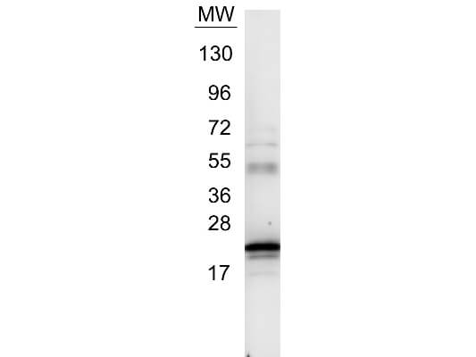
Western blot using Rockland's Anti IL6 antibody. Protein was resolved on a 4 20% Tris Glycine gel by SDS PAGE and transferred onto nitrocellulose. The blot shows detection of a band ~21 kDa in size corresponding to anti IL6 antibody. Molecular weight markers are also shown (MW). After transfer, the membrane was blocked for 30 minutes with 1% BSA TBST. Detection occurred using peroxidase conjugated Goat anti Rabbit IgG (p/n 611 103 122) secondary antibody diluted 1:40,000 in blocking buffer (p/n MB 070) for 30 min at RT followed by reaction with FemtoMaxTM chemiluminescent substrate. Image was captured using VersaDocTM MP 4000 imaging system (Bio Rad).
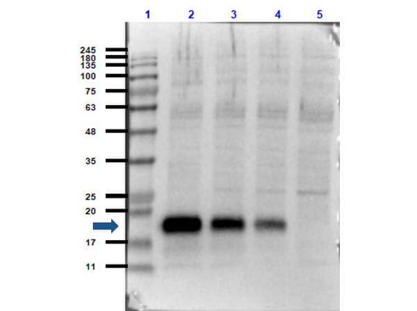
Western Blot of Rabbit Anti-IL-6 Antibody. Lane 1: Opal Prestained Molecular Weight Marker (p/n MB-210-0500). Lane 2: Human Rec. IL-6/HeLa Whole Cell Lysate [0.05ug/10ug]. Lane 3: Human Rec. IL-6/HeLa Whole Cell Lysate [0.02ug/10ug]. Lane 4: Human Rec. IL-6/HeLa Whole Cell Lysate [0.01ug/10ug]. Lane 5: HeLa Whole Cell Lysate (p/n W09-000-364) [10ug]. Primary Antibody: Anti-IL-6 at 1:1000 overnight at 2-8C. Secondary Antibody: Goat Anti-Rabbit IgG HRP conjugate (p/n 611-103-122) at 1:70,000 for 30mins at RT. Block: BlockOut Buffer (p/n MB-073). Predicted MW: ~23kDa. Observed MW: ~19 Exposure: 2 seconds.
|
|
|
|
Western blot using Rockland's Anti IL6 antibody. Protein was resolved on a 4 20% Tris Glycine gel by SDS PAGE and transferred onto nitrocellulose. The blot shows detection of a band ~21 kDa in size corresponding to anti IL6 antibody. Molecular weight markers are also shown (MW). After transfer, the membrane was blocked for 30 minutes with 1% BSA TBST. Detection occurred using peroxidase conjugated Goat anti Rabbit IgG (p/n 611 103 122) secondary antibody diluted 1:40,000 in blocking buffer (p/n MB 070) for 30 min at RT followed by reaction with FemtoMaxTM chemiluminescent substrate. Image was captured using VersaDocTM MP 4000 imaging system (Bio Rad).
|
|
|
| メーカー |
品番 |
包装 |
|
RKL
|
109-401-310
|
1.0 ML
|
※表示価格について
| 当社在庫 |
なし
|
| 納期目安 |
約10日
|
| 保存温度 |
-20℃
|
|

